
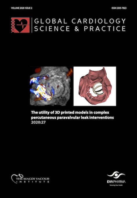
The utility of 3D printed models in complex percutaneous paravalvular leak interventions
DOI:
https://doi.org/10.21542/gcsp.2020.27Abstract
Paravalvular leaks (PVL) are seen in 5-17% of patients after surgical mitral and aortic valve replacement. This is usually well-tolerated in the majority of patients; however, up to 5% will require re-intervention due to either hemodynamically significant regurgitation or hemolysis requiring repeated blood transfusion. Transcatheter closure of PVLs is becoming the treatment of choice in many patients owing to the high risk of redo surgery, high rates of recurrence with the surgical approach, and substantial improvements in device technology and growing expertise in structural heart disease interventions. Careful selection of the appropriate candidates by the Heart Team with in-depth analysis of clinical and multimodality imaging data is critical to ensuring good short- and long-term outcomes.
The defect is usually oval/ crescentic and often serpiginous in nature, which poses significant challenges in the optimal size and number of devices to implant - especially with large size defects. Generally, defects involving more than 25-30% of the sewing ring are generally deemed unsuitable for percutaneous closure. While the Amplatzer family of vascular plugs (e.g. AVP3 and AVP2) is commonly used for percutaneous closure of PVLs, there are currently no approved dedicated devices for this indication, except the paravalvular leak device (Occlutech) which is not universally available. Small and relatively circular defects can usually be closed using a single plug, conventionally utilizing a size that is 25-30% larger than the mean diameter of the defect. Larger and crescentic defects on the other hand frequently require more than one plug and can be quite challenging in terms of choosing the appropriate size(s).
We report two cases with very large defects with irregular shape in which 3D printed modeling was extremely useful for bench testing to optimize the number and sizes of devices to be implanted.
Downloads
Published
Issue
Section
License
This is an open access article distributed under the terms of the Creative Commons Attribution license CC BY 4.0, which permits unrestricted use, distribution and reproduction in any medium, provided the original work is properly cited.
