
Intracoronary near-infrared spectroscopy: an overview of the technology, histologic validation, and clinical applications
DOI:
https://doi.org/10.21542/gcsp.2016.18Abstract
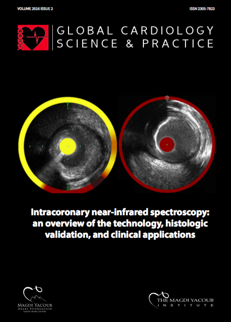
Intracoronary near-infrared spectroscopy (NIRS) imaging, which is now clinically available in a combined NIRS and intravascular ultrasound catheter, is a novel catheter-based imaging modality capable of identifying lipid core plaque within the coronary arteries of living patients. The present manuscript provides an overview of intracoronary NIRS imaging with a focus on several concepts essential to individuals seeking to better understand this novel imaging modality. One of the major assets of NIRS is that it has been rigorously validated against the gold standard of histopathology and has been shown to accurately identify histologically-proven fibroatheroma. Clinical studies of NIRS have demonstrated its ability to accurately identify large lipid core plaques at culprit lesions across the spectrum of acute coronary syndromes. NIRS has also been shown to detect lesions at increased risk of causing peri-procedural myocardial infarction during PCI. With regards to predicting future risk, NIRS is seemingly capable of identifying vulnerable patients at increased risk of experiencing subsequent patient-level cardiovascular events. In addition to these clinical applications of NIRS, there are several large prospective observational studies underway to determine if NIRS imaging will be able to identify vulnerable plaques at increased risk of triggering site-specific future coronary events. These studies, once completed, are anticipated to provide valuable data regarding the ability of NIRS imaging to identify plaque vulnerability.
Downloads
Published
Issue
Section
License
This is an open access article distributed under the terms of the Creative Commons Attribution license CC BY 4.0, which permits unrestricted use, distribution and reproduction in any medium, provided the original work is properly cited.
